The 5 medical highlights of 2020


The turbulent year has passed, health became the most concern and medical technologies have been successfully developed all around the world. Among them, the deep learning application in MRI scanners, artificial skin, restoring sight to those with lost vision; testing antibiotic resistant strains and finding the structure of protein to help influenza B are the most outstanding technologies of 2020.
Changing the Game In Magnetic Resonance Imaging
The application, AIR™ Recon DL* runs on GE’s Edison™ software platform. It uses a deep learning algorithm to improve MR image reconstruction. For radiologists, this translates as sharper images in a shorter amount of time. And the quicker doctors can come up with diagnoses, the faster patients can start treatment.
MRI scanners utilize powerful magnets — hidden inside the large donut surrounding the patient — to excite hydrogen atoms (protons) in the body. Those hydrogen protons then emit radio signals, which are detected by the scanner and transformed into a digital image. But those radio signals compete with other radio waves generated by the MRI apparatus and the human body itself — a kind of ambient electrical noise.
At Michigan State University, researchers created the first “miniature human heart model” in the lab; the small organoid contains “all primary heart cell types and a functioning structure of chambers and vascular tissue.”
The lab-grown organs — called human heart organoids, or HHOs — could offer researchers new insights into the genesis of disorders that develop in fetal hearts. (Roughly 1% of all live births involve congenital heart defects, the most common birth defect in humans.) Previously, they’ve been limited to animal models, donated fetal remains and in vitro cell research. “Now we can have the best of both worlds, a precise human model to study these diseases — a tiny human heart — without using fetal material or violating ethical principles,” said biomedical engineering professor Aitor Aguirre, the senior author of a paper on the organoids published on the preprint server bioRxiv. “This constitutes a great step forward.”
The creation of HHOs relies on induced pluripotent stem cells, a kind of undifferentiated cell that can be directed to grow into any kind of cell in the body. Aguirre explained, “This process allows the stem cells to develop, basically as they would in an embryo, into the various cell types and structures present in the heart. We give the cells the instructions and they know what they have to do when all the appropriate conditions are met.”

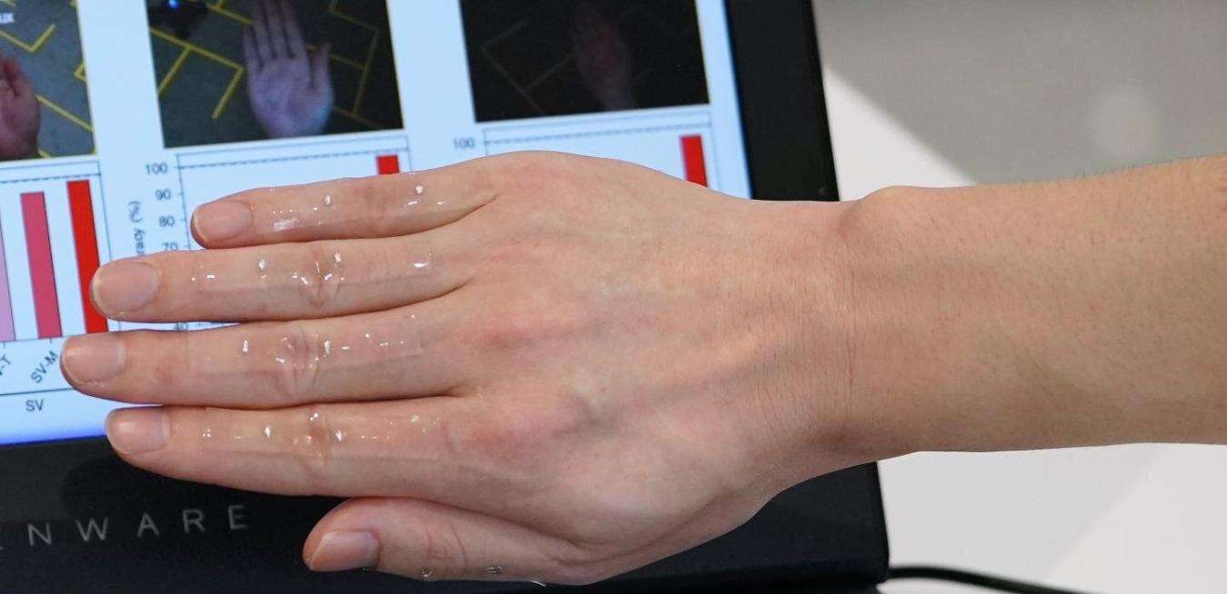
The team fabricated a transparent, stretchable strain sensor that adheres to the skin but cannot be seen in camera images. Caption and image credit: NTU Singapore.
What is it? Scientists at Singapore’s Nanyang Technological University combined “skin-like electronics with computer vision” into an artificial intelligence system that can recognize hand gestures.
Why does it matter? The technology could have uses in surgical robots, gaming interfaces, and robot-aided workplaces. Markus Antonietti, the director of Germany’s Max Planck Institute of Colloids and Interfaces — who was not involved in the project — said in NTU’s press release that “the findings from this paper bring us another step forward to a smarter and more machine-supported world. Much like the invention of the smartphone, which has revolutionized society, this work gives us hope that we could one day physically control all of our surrounding world with great reliability and precision through a gesture.” The paper was published in Nature Electronics.
How does it work? The Singaporean team’s “bio-inspired” system includes a stretchable sensor, made of single-walled carbon nanotubes, that fits over the hand, while the AI system combines three different neural network approaches: one concerning visual processing, one concerning somatosensory processing and one that fuses the two. NTU’s Chen Xiaodong, the study’s lead author, said the technology is “unique” in that it resembles “the somatosensory-visual fusion hierarchy in the brain.”

Various components of the test for antibiotic-resistant bacteria developed by BYU researchers. Image credit: Claire Moore/BYU.
What is it? An interdisciplinary team from Brigham Young University developed a test that can detect the presence of antibiotic-resistant bacteria in the blood in less than an hour.
Why does it matter? On a broad scale, antibiotic-resistant bacteria are a major health crisis. For patients, they’re a severe health threat that advances quickly in the body and can have lethal effects. Current testing methods can take up to 24 hours or longer, though, to return results. "Once you’re trying to diagnose the disease, the clock is ticking," said Aaron Hawkins, a professor of electrical and computer engineering and the co-author of a new study in the journal Lab on a Chip. “Every hour the disease is untreated, survivability drops by about 7%. You want to know what you’re fighting immediately so you can apply the right treatments.”
How does it work? As that journal name indicates, the method involves a chip. First researchers take a blood sample and spin it to isolate the bacteria, then DNA is extracted from the bacteria. The DNA is pushed through a fluid channel on a microchip, where a fluorescent signal will indicate the presence of antibiotic-resistant strains. Importantly, the chip can test for the presence of multiple resistant bacteria at once. The researchers plan to install the chip on a disposable, inexpensive cartridge that can be used in hospitals.

A new protein could help researchers understand how strains of the flu virus differ, and develop drugs to combat them. Image credit: Getty Images.
What is it? Chemists at the Massachusetts Institute of Technology “discovered the structure of a key influenza protein” called BM2.
Why does it matter? There are three classes of flu virus — A, B and C — with A usually being the most common, especially early in flu season. This year, though, influenza B has predominated, accounting for about two-thirds of reported cases in the U.S. Each class of flu virus is associated with a different kind of M2 protein, and getting a snapshot of its structure in flu B — which is what the MIT team focused on, using nuclear magnetic resonance spectroscopy — could help researchers understand how the virus strains differ, and develop drugs to combat them.
How does it work? BM2 is a “proton channel that controls acidity within the virus,” per MIT, helping it release genetic materials into infected cells. Mei Hong, an MIT chemistry professor and senior author of a new paper in Nature Structural & Molecular Biology, said, “If you can block this proton channel, you have a way to inhibit influenza infection. Having the atomic-resolution structure for this protein is exactly what medicinal chemists and pharmaceutical scientists need to start designing small molecules that can block it.”