Medical Imaging
These Machines Helped Unveil Secrets Of The Human Body


 Elihu Thomson’s X-ray machine from 1896. Image Credit: GE. Top Image: The latest GE imaging systems like the Revolution CT can produce detailed images of the vascular system. Image Credit: GE Healthcare.
Today, GE Healthcare makes everything from advanced imaging machines to super-resolution microscopes and software that can be used to process, analyze and probe terabytes of data produced by the machines for insights. The business generated $17.6 billion in revenues in 2015.
 Above: An MRI image of the brain. The method, called diffusion tractography, is displaying some of the long white matter bundles (red: left-right, green: anterior-posterior, blue: head-foot). Top image: The skull and carotid arteries imaged by GE's Revolution CT Scanner. Images credit: GE Healthcare
Some of the technology was on display at the 101th annual meeting of the Radiological Society of North America (RSNA) in Chicago last December. The RSNA is the radiological industry’s “Grand Slam” gathering and trade show that was expected to draw 60,000 visitors and exhibitors this year. GE attended the inaugural meeting in 1914 .
 An Edison X-ray ad from 1897. Image credit: GE
In 2015 at RSNA, GE announced the first GE Health Cloud that will initially gather data from 500,000 GE imaging machines, allow doctors to collaborate online, and let independent software vendors to develop apps in the new cloud ecosystem. The GE machines that will supply the data draw on decades of research and commercial development starting with Thomson’s fluoroscope, the world’s first commercially available X-ray machine.
 GE's William Coolidge invented what is considered the modern X-ray tube. He also developed an early portable X-ray machine. Coolidge's X-ray machine was used in military hospitals during World War I. Image credit: Museum of Innovation and Science Schenectady
In 1932, GE’s Irving Langmuir won the Nobel Prize in Chemistry for his work that led to early coronary artery imaging. In 1973, his colleague Ivar Giaever received the Nobel Prize in Physics for research that led to the first GE MRI machine a decade later. In the 1980s their colleague John Schenck at GE Global Research took the first brain selfie with a GE MRI scanner. The list of innovations goes on. Take a walk with us from the past into the future.
 General Electric researcher and scientist Irving Langmuir receives the 1932 Nobel Prize in Chemistry in Stockholm, Sweden. Langmuir was associate director of the GE Research Laboratory at the time. Image credit: Museum of Innovation and Science Schenectady
 Dr. Ivar Giaever, 1972 recipient of the Nobel Prize in Physics, poses with his superconductive tunneling experiment. Image credit: Museum of Innovation and Science Schenectady
 The Nobel acceptance telegram from Ivar Giaever. Image credit: Museum of Innovation and Science Schenectady
 In 1939, GE medical scanners produced X-ray images of mummies for the New York World’s Fair. Scientists are still using GE imaging machines to study ancient objects, including mummies, a baby woolly mammoth and a piece of a sunken ship. Image courtesy of the New York Public Library.
 John Schenck's work is featured in Breakthrough, the six-part science TV series developed by GE and National Geographic Channel. He took the first brain selfie with a GE MRI scanner. Image credit : GE Reports
 This image shows complex patterns of connectivity of the human cortex measured in vivo with MRI via diffusion of water molecules in axons in the white matter. Image credit: GE Global Research
 A 3D view of the human heart’s mitral valve — two leaflets that open and close with each heartbeat. The mitral valve ensures that blood flows in one direction only. The image was taken with "4D" ultrasound. Image credit: GE Healthcare
 The pair of “bubbles” in this image are actually a twin pregnancy. Each of the amniotic sacs has a 6-week-old embryo inside. The image was taken with "4D" ultrasound. Image credit: GE Healthcare
 An image of the body with GE's Revolution CT scanner. Image credit: GE Healthcare
 An image of the heart with GE's Revolution CT scanner. Image credit: GE Healthcare
 An MR image of the liver. A 16 year old obese patient with elevated liver enzymes and fatty liver infiltration on ultrasound and MR. The MR elastography (MR Touch) was performed to evaluate tissue stiffness prior to a planned biopsy. The MRE showed normal liver stiffness, indicating the presence of simple steatosis, but no fibrosis or inflammation. The biopsy was cancelled. Image credit: GE Healthcare
 Try finding and untying single hairs within a tangled knot. FlightPlan for liver software makes identifying tumor-feeding vessels easier by highlighting the cancer-feeding vessels with high sensitivity. Image credit: GE Healthcare
 ViosWorks* combines 3D cardiac anatomy, function, and flow in 1 free-breathing, approximate 8 minute scan. It enables visualization of the whole chest and beating heart from any vantage point – any structure, in any plane – simultaneously seeing ventricles contracting and accurately quantifying blood flow. *7 dimensional viewing capabilities of the heart; 3 in space, 1 in time, 3 in velocity direction. Image credit: GE Healthcare
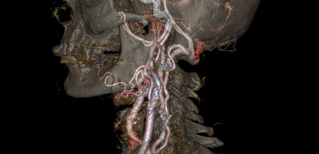
 The skull and carotid arteries captured by GE's Revolution CT scanner. Image credit: GE Healthcare.
You can find more images on GE Healthcare's Pulse site.