Medical Imaging
Little Wonders: Neonatal Surgeon Captures Stunning Images With 4D Ultrasound


Shih, who serves as the head of the labor ward and as an assistant professor in the department of obstetrics and gynecology at National Taiwan University Hospital, scanned the mother with an ultrasound that allowed him to see clearly inside the womb and understand the twins’ location. He decided to perform a cesarean section the next day because the risk of preterm birth was less than fetal surgery.
Then, during the operation, something unexpected happened. “The hand of one twin suddenly extended outside the uterine cavity and tensely grasped my finger like a drowning person grasping a floating piece of wood,” Shih told GE Reports last year. “It looked like a call for help to me. Everyone here held their breath. Although nobody taught the little baby, she presented a strong wish to survive. I felt touched. It’s a moment I will never forget.” The surgeon has kept the image to remind him of the human will to survive.
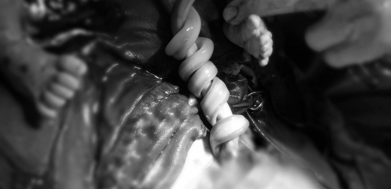
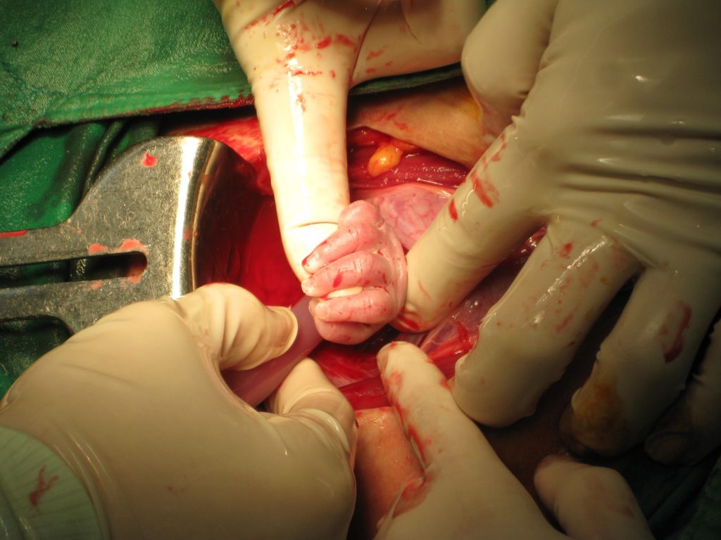 Top image: Because monoamniotic twins live in the same “room,” there’s enough fluid space for them to swim around and even hug each other. The danger is that the umbilical cords may wrap around the babies. Above: “The hand of one twin suddenly extended outside the uterine cavity and tensely grasped my finger like a drowning person grasping a floating piece of wood,” Dr. Shih said. Images credit: National Taiwan University Hospital
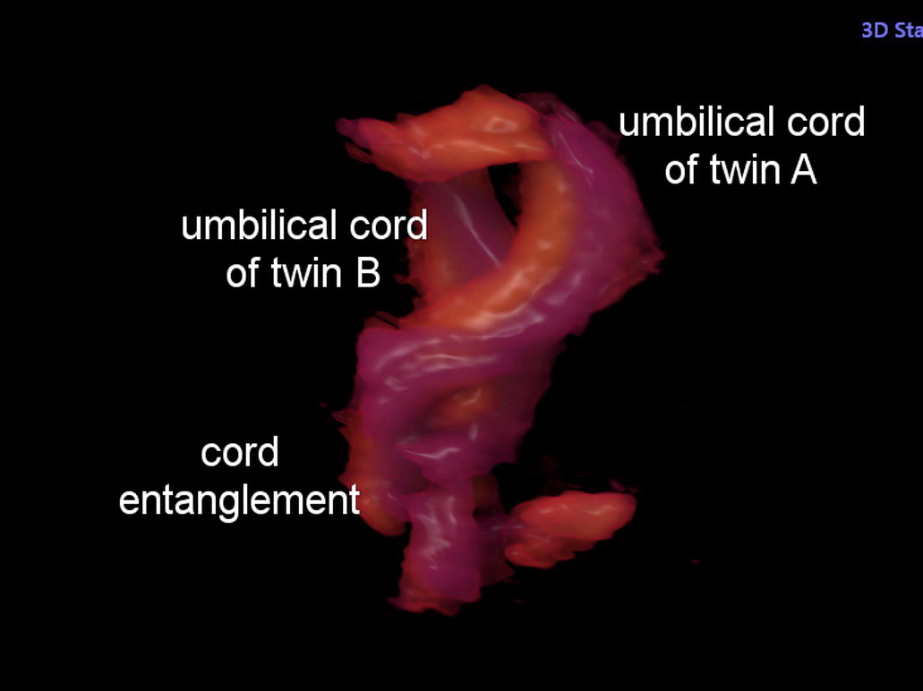
Shih is also the secretary general of Taiwan’s Society of Ultrasound in Medicine and was an early adopter of 4D ultrasound in 2002. This technology, which can monitor the fetus in the mother’s belly with startling clarity in the three dimensions and over time, has enabled him to snap other breathtaking images, including a picture of an entangled umbilical cord he took while performing a C-section on monoamniotic twins — identical twins who share an amniotic sac within their mother’s uterus.
Because monoamniotic twins live in the same “room,” there’s enough fluid space for them to swim around and even hug each other. The danger is that the umbilical cords may wrap around the babies, strangling or suffocating them.
These twins’ mom had visited Shih because one of the fetuses had a complex heart condition that would require surgery after birth. Using a 4D ultrasound system called Voluson E10, Shih found that the twins had a cord entanglement, which typically requires an early delivery. However, the minimum weight for open-heart surgery is 2.5 kilograms (about 5.5 pounds), and in this case the twin was too small.
Shih continued to monitor the twins with ultrasound and found the cord entanglement was not severe. The parents decided to deliver the babies closer to the due date so that the smaller baby had time to grow in strength and size.
“This finding encouraged me to wait for more time to allow the baby to grow inside the womb,” Shih explained. “Ultrasound is incredibly helpful in these situations because it can depict the cord entanglements and alert the clinician if an earlier delivery is necessary.”
Shih delivered the babies via C-section at 35 weeks. The baby requiring open-heart surgery recovered successfully, and today the twins are 4 months old and are doing well. So are their peers Shih delivered two years earlier.
The doctor is wistful about his experiences in the operating room. The day before the planned C-section on the twins with the entangled umbilical cords, Shih spotted something he had never seen before: The twins’ fetal hearts were beating at the exact same pace for several hours. “It essentially disobeys the textbook description,” he says. “We are taught that if you record the same heartbeat for twins, you are catching the same fetus. However, we checked the ultrasound again and again, and proved there was a concordant cardiac rhythm coming from both hearts. It’s something we still cannot explain.”
A version of this article originally appeared on The Pulse, GE Healthcare’s newsroom.