Nadeem Ishaque: Imaging the Brain in Real-Time


New tools are needed to study the brain at multiple scales from the molecular and cellular level to the level of circuits and networks and, finally, to the level of overall anatomic and functional organization. Such understanding is vital in developing novel treatment and diagnostic strategies for debilitating neurodegenerative and neuropsychiatric disorders for which no cure exists today.
The Obama administration, through The National Institutes of Health’s “BRAIN” (Brain Research through Advancing Innovative Neurotechnologies) initiative, is dedicating resources to enable leading academic and industrial research institutions to take non-incremental, major steps forward in brain research through a competitive, peer-reviewed process.
One of these exciting “major step forward” projects – which GE is embarking on with partners from the West Virginia University, the University of Washington and the University of California-Davis — is to make imaging of the brain in action, in real-time, a possibility.
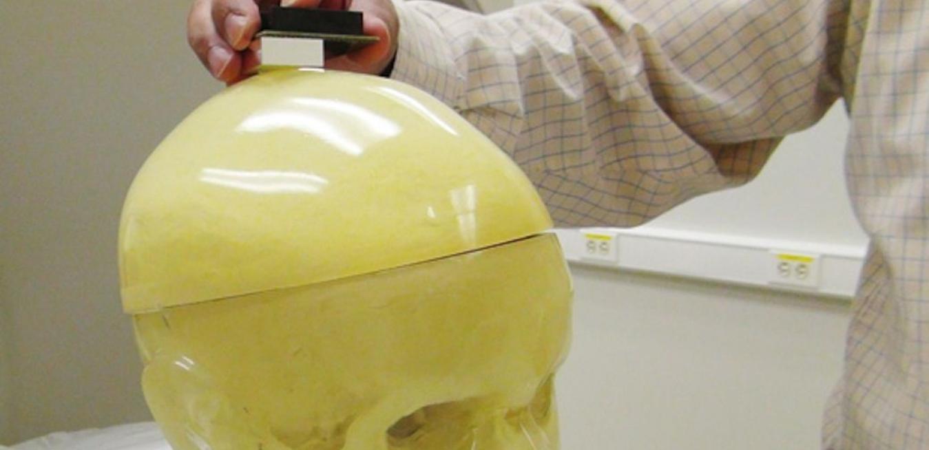
If successful, this effort would represent a monumental advancement in imaging technology that would enhance the understanding of brain function both in normal and disease states. Today, medical professionals and researchers do not have the ability to image subjects unless they are lying down in a resting state. GE scientists are beginning to investigate ways to make a wearable imaging device for the brain. This helmet would be lined with positron emission tomography (PET) detectors several times smaller and thinner than those found in a standard, whole body PET scanner, and would allow for a patient to be imaged in a more natural setting — upright and mobile
In this video from our upstate N.Y. labs, my colleague Ravi Manjeshwar demonstrates some of the technology breakthroughs we’ve already achieved and outlines where the project is headed next:
The high-sensitivity, high-resolution approaches that we plan to utilize in this PET imaging system will allow physicians to image cells and misfolded proteins in the brain that characterize neurological disorders. Today, many important classes on neurons and glial cells remain undetectable by imaging techniques because of their very low concentrations. The concept we are developing aims to detect biomolecules in as little as femto-molar concentrations. This will open up new fields of tracer development and molecular imaging research to better understand the integration points in key signaling networks and assess biologically important targets.
Additionally, the micro-dose concept we’re exploring would reduce radiation exposure to patients to only about as much radiation as a cross-country flight, while still delivering high-resolution images. Such negligible radiation exposure is crucial in allowing otherwise healthy subjects to undergo PET brain scans with this new system.
Working with the academic community, we hope to use our technologies to help close the gap in understanding of the brain.
Nadeem Ishaque is Global Technology Director of Diagnostics & Biomedical Technologies at GE Global Research.