Here’s Looking at You, Kid: Get to Know Your Baby Before It's Born With 4D Ultrasound


Working in the late 1700s, Spallanzani showed that blindfolded bats could still catch flies and find their way around. But they failed miserably when he sealed up their ears. His discovery of the bat’s “sixth sense,” called echolocation, launched the science of ultrasound.
The pair of “bubbles” in this image are actually a twin pregnancy. Each of the amniotic sacs has a 6-week-old embryo inside. Top image: A real-time 4D ultrasound GIF of a baby’s face.
It took scientists more than a century to explain Spallanzani's mysterious results and build machines that, like the bat’s larynx and ears, generate inaudible noise and then analyze its echoes. During World War I, the Royal Navy used an early, man-made version of the system to find and sink a German U-Boat in the Atlantic. But things have gotten much more refined since.
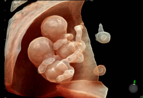
Another image of a twin pregnancy at 8.5 weeks.
Today, the latest applications of the technology include “4D" ultrasound machines. They can monitor the fetus in the mother’s belly with startling clarity in the three dimensions and over time. “In the past, you could see a flat two-dimensional image of the fetal profile,“ says Barbara Del Prince, a global managing director for ultrasound products at GE Healthcare. "But today you can watch their movements in 3D, see a smile or a grimace, glimpse their personality.”
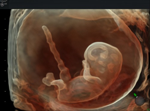
This image of a 9-week-old fetus shows developing brain structures.
Parents love seeing the pictures, but doctors use the technology to study organs and functions of the fetus like the structure of the brain (see image above) and the working of the heart (below).
Del Prince says that GE’s most advanced system, Voluson E10*, can emit signals and process information fast enough to view the heart in real time. “It may help doctors to make confident diagnoses sooner,” she says.
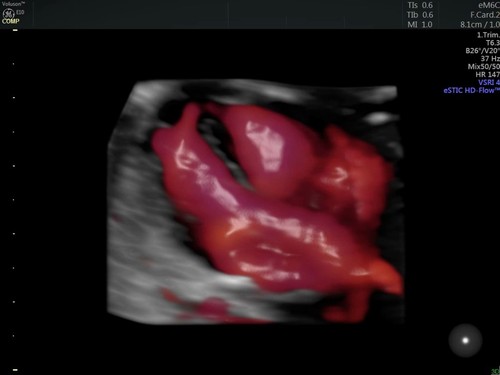
An image of the developing heart taken during the first trimester with HDlive Flow.
The system is so fast because it is equipped with a new probe technology called “Electronic 4D.” The probe is using more than 8,000 piezoelectric crystals to electronically steer the ultrasound beam and provide clarity and speed.
A still image of a foot captured from 4D ultrasound with HDlive Silhouette.
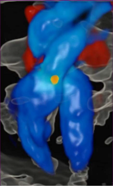
Another software feature, HDlive Flow, generates a 3D-view of the blood flow and provides a realistic rendering of blood vessels. “This is really important for doctors when they are looking at abnormalities,” Del Prince says. “But most parents will probably remember the first smile.”
The Voluson E10 is currently available in the U.S., Europe and Japan.
*Voluson is a trademark of the General Electric Co. or one of its subsidiaries.