The final frontier


A new generation of high powered microscopes is making it possible not only to see, but look inside these tiny predators, gaining vital knowledge which will one day help to defeat existing pathogens as well as emerging antibiotic resistant strains of disease.
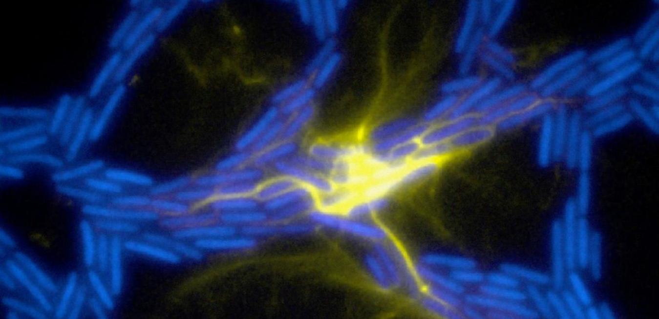
This new breed of microscope can see single objects that are 100 nanometres in size. This allows scientists to see the little individual fibres that propel bacteria across surfaces, and to observe where proteins they produce move within a single bacterial cell. The emergence of OMX microscopy technology, developed by GE Healthcare, has allowed a level of detail to be observed in three dimensions that was not possible in previous traditional microscopes.
Unlike traditional microscopes, OMX microscopes have no ocular lens. Samples are placed on a platform within the machine, photographed by multiple high resolution cameras and projected onto a computer screen. The microscopic images are captured using an ultrafast structured illumination module, enabling researchers to perceive the shape and observe the movement of these miniscule monsters. The results are highly detailed images which display samples in three dimensions, capturing intracellular elements which through traditional microscopes would be no more than indistinct blurs.
“We’ve already been able to look inside the malaria parasite, and watch it change over time, we can see how it reproduces and causes the red blood cell to become misshapen,” said Associate Professor Cynthia Whitchurch from the Microbial Imaging Facility at the University of Technology Sydney.
“We’ve also been able to see that the muscle proteins in patients with Muscular Dystrophy don’t go to the right positions inside the cell after a wound occurs when compared to what normal cells do, because with this technology we can actually see where these proteins travel inside each cell in unprecedented detail."
Now the second generation OMX Blaze microscopes allow samples to be captured at the rate of one image a second, creating time-lapse sequences from live samples.
According to UTS Research Fellow, Dr Lynne Turnbull, Australia’s foremost expert in the use of the OMX technology, this ability to capture movement will lead to some dramatic breakthroughs in microbiology.
“Before OMX we could see that there were different proteins in different locations inside a bacterial cell but not in much detail,” said Dr Turnbull. “Now we are able to see how these proteins within a bacteria change position over time or how one protein might change its shape over time, such as when the cell divides, leading to a new model of cell division based on this evidence.”
“This technology will be fundamental to research into some of the most serious threats we currently face, including antibiotic resistance in bacteria,” Associate Professor Whitchurch added.