×

GE.com has been updated to serve our three go-forward companies.

Please visit these standalone sites for more information

GE Aerospace | GE Vernova | GE HealthCare 

header-image

Slime Science: The “OMG Microscope” Helps Scientists See How Bacteria Creep

July 16, 2013
Australian scientists have used a powerful GE microscope to study the spread of slimy, drug-resistant bacterial colonies called biofilms. They have been able to determine how the microbes stick together and move in intricate, self-organized patterns. The research could help doctors fight aggressive infections caused by biofilms colonizing catheters and other medical devices. “Biofilms are notoriously difficult to clear,” says team leader Cynthia Whitchurch, senior research fellow at University of Technology Sydney. “The bacteria can actively migrate along the surface and move up to the bladder and the kidneys, where they can cause a lot of problems. We are trying to understand how the bacteria coordinate to be able to achieve this migration so we can come up with smart ways to inhibit that.”
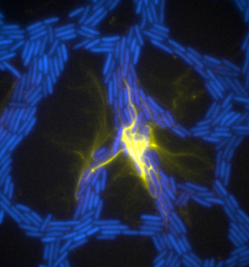
The team used a number of imaging technologies including GE’s DeltaVision OMX Blaze microscope equipped with high-speed cameras. They observed for the first time how bits of DNA that stick to the outside wall of the bacteria hold the biofilm together and guide its movement. Scientists call this microbe glue extracellular DNA, or eDNA. “We knew it was there - we had evidence from observing how the bacteria were behaving - but we just couldn’t see it or detect it in any way,” Whitchurch says. “The high-sensitivity cameras on the OMX Blaze enabled us to answer the question of how the bacteria were gluing themselves together.”

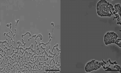
The researchers released their findings in the current issue of the Proceedings of the National Academy of Sciences. They observed cell movements during the expansion of living biofilms by combining microscope images with sophisticated computer-vision algorithms. Their analysis found “highly coherent groups of bacteria” at the leading edges of the biofilm. These groups create channels that help cells following behind them migrate, and leave eDNA that “facilitates efficient traffic flow” throughout the channel network.

 /><br />
<br />
<em> Extracellular DNA (yellow) in biofilms of the bacterial pathogen Pseudomonas aeruginosa organizes traffic flow of individual bacteria (blue) as they move through the biofilm trail network. Credit: E. Gloag and L. Turnbull, The ithree institute, University of Technology Sydney.</em><br />
<br />
This insight could guide scientists to new ways of fighting biofilms. “We could introduce artificial channels along the surface of the device and confuse the bacteria so they can’t be as effective,” Whitchurch says. “Another option would be destroying the eDNA.”<br />
<br />
Scientists have recently used the OMX microscope to observe malaria parasites attacking blood cells, the response of cancer cells to chemotherapy, and even the cell-to-cell transmission of HIV. Jane Stout, a research associate at Indiana University, used it to study cell division and dubbed the machine the “OMG.” Dr. <a href=Francis Collins, director of the National Institutes of Health, agreed. He called images produced by the OMX “showstoppers.”

Delta Vision OMX is for research use only, not for use in diagnostic procedures. Delta Vision OMX is a GE trademark.

 /><br />
<br />
<em>Pseudomonas aeruginosa is a bacterial pathogen that is able to <a href=actively colonize surfaces via a process called twitching motility.