Medical Imaging
Puppy Love: Spanish Vets Are Using Innovative MRI Techniques To Unravel The Mysteries Of Canine Epilepsy


Canine epilepsy might be a fairly common neurological affliction, affecting around 1 in every 25 dogs,[1] but its effects are distressing for both the pet and its owner. Until recently, the condition was also something of a mystery. Head trauma, brain tumors and cerebrovascular disease can all trigger seizures, as can idiopathic epilepsy, which is a genetic disorder.
But times are changing. Today’s dog owners pay more attention to their pooches’ well-being and are more likely to take their beloved pets to a veterinary hospital for tests and treatment after they suffer seizures. And vets are increasingly using state-of-the-art magnetic resonance imaging (MRI) scanners and techniques that are common in human medicine to diagnose and treat canine epilepsy, as well as the other neurological diseases and injuries afflicting “man’s best friend.”
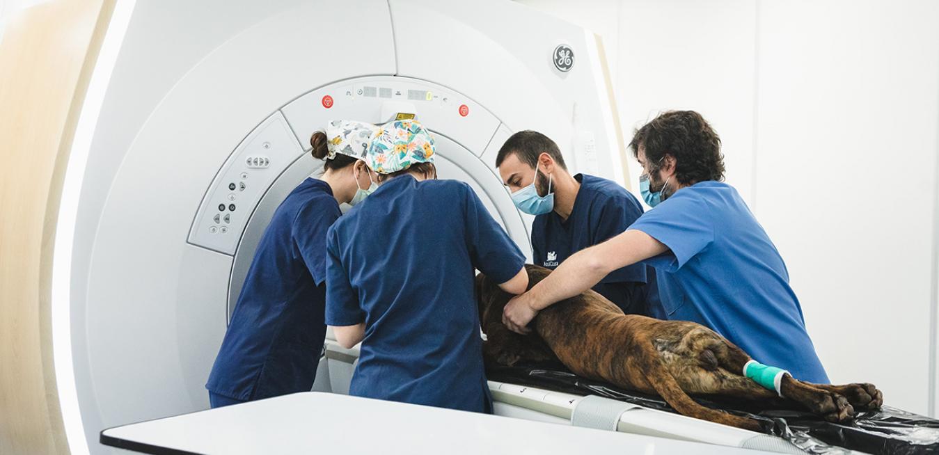
Take the Anicura Valencia Sur Veterinary Hospital,[2] a facility on Spain’s Mediterranean coast that treats around 5,000 animals per year. Anicura’s vets are using a GE Healthcare-made MRI machine to image dogs’ brains and spinal cords to help diagnose and treat the tumors, strokes and other disorders that trigger epileptic episodes. The device is a cut above the conventional MRI scanners used in many Spanish veterinary facilities because it boasts more powerful magnets and newer software features. This opens up a world of new MRI techniques for vets, who can now optimize the sequence and strength of magnetic pulses to produce crisper images more quickly.
In fact, you would see exactly the same scanners and techniques in the radiology department of any of Valencia’s major hospitals that treat humans. Sergio Ródenas, a neurological specialist at Anicura, says the combination of superior MRI scanners and software with innovative techniques will help to drive better outcomes for Valencia’s cherished dogs. “They are allowing for early diagnosis in a way that we could never imagine before.”
If you have ever taken a sickly dog or cat to the vet, you’ll know that their first port of call is often the radiology department. “We’ll image and diagnose animals in the same way that we do for humans,” says Ródenas. He reels off a list: “X-ray, ultrasound, computerized tomography or MRI.” In human medicine, doctors use MRIs to image a wide range of anatomy: the brain and spinal cord, bones and joints, breasts, hearts and blood vessels, and internal organs. But vets have traditionally used MRIs more sparingly, partly because the exams can be expensive. “Probably up to 90% to 95% of MRI exams in veterinary medicine are brain or spinal cord scans,” says Ródenas.
Anicura’s vets generally sedate a dog before an MRI exam to ensure the pup stays still inside the scanner’s donut-shaped hole. Fortunately, the scans don’t take long. The device at Anicura boasts more powerful magnets than conventional MRI machines in the veterinary market, and they create an ultra-stable magnetic field inside the donut hole. That rock-solid field holds fast the positively charged particles called protons in the hydrogen atoms of the pooch’s body. The scanner’s computer then produces detailed MRI images from the millions of data points collected: The variations in energy across the hydrogen atoms. “The quality of images [from the high-field scanner] is vastly superior [to conventional MRI machines],” explains Ródenas.
The fine contrast in the resulting grayscale images allows vets to quickly zero in on the telltale signs of common brain and spinal cord diseases and injuries, including tumors, suspicious inflammation and ischemic disorders. Ródenas says that some of these pathologies, all of which can trigger seizures, often go unnoticed in CT or X-ray exams, as MRI is best positioned to image soft tissue and neurological structures.
Other MRI techniques involve varying the strength, duration and angle of magnetic pulses. “None of these are invasive,” says Ródenas, “they just allow for early diagnosis.” Among those techniques: Perfusion-weighted MRI, which gives vets insight into how blood is moving through tissues; MR spectroscopy, which images tissue to identify metabolites, possibly indicating the presence and type of a tumor; and diffusion MRI, which measures the random motion of water molecules in tissue, allowing vets to improve stroke diagnosis or better characterized tumors.
The techniques boost clinical confidence in tumor diagnosis, says Ródenas. “We can then [recommend] doing a biopsy or surgery.” The Valencian vets can also analyze images to ascertain whether a dog has suffered a stroke, allowing vets to give pet owners a clearer prognosis.
Of course, the MRI techniques might not reveal any pathology at all, in which case vets might diagnose idiopathic epilepsy and prescribe a course of anti-epileptic medication for the animal. This diagnosis may not comfort the dog owner, but provides assurance that the owner is doing everything possible.
Vets can also use the devices to help diagnose musculoskeletal diseases or other spine disorders, such as spinal cord embolisms, inflammation and contusions. Ródenas says that vets welcome every last pixel of detail because the wide variety of patients and diseases can complicate their jobs. The human species doesn’t have much genetic variation, but there are hundreds of breeds in the dog kingdom. “Dogs may range from 1 kilogram to 80,” says Ródenas.
These techniques aren’t reserved for dogs; they can also help image cats. Cats’ differential diagnoses are not the same as dogs’; they are more prone to encephalitis (brain inflammation) and brain tumors, explains Ródenas. The vet says he has even carried out MRI scans on rabbits. “They can suffer otitis [ear inflammation], encephalitis or cerebrovascular issues,” he explains.
Ródenas hopes that MRI scanners and techniques will become a common sight at vet clinics in Spain, where pet ownership has soared in recent years.[3] “[MRI] use in Spain is not very widespread due to the lack of information,” he explains. “But many people have tremendous love for their pets.”
Top: Veterinarians at Anicura Valencia Sur Veterinary Hospital in Spain are using a GE Healthcare-made MRI machine to image dogs’ brains and spinal cords to help diagnose and treat the tumors, strokes and other disorders that trigger epileptic episodes. Image credit: GE Healthcare.
A version of this story originally appeared on GE Healthcare's Insights blog.
Dr. Sergio Ródenas has no contractual relationship with GE Healthcare beyond being an end user of a GE medical device.
The statements described by GE's customer are based on their own opinions and on results that were achieved in the customer's unique setting. No diagnostic statements are inferred or included in these materials. All clinical diagnosis should be made by a trained physician or clinician.
[1] Seizures and epilepsy in dogs, https://www.bluecross.org.uk/pet-advice/epilepsy-and-seizures-dogs.
[2] Anicura Hospital Veterinario Valencia, Sur https://hvvalenciasur.com/.
[3] With more Spaniards living alone, pet numbers soar in the cities: https://english.elpais.com/elpais/2019/05/22/inenglish/1558512182_011864.html.