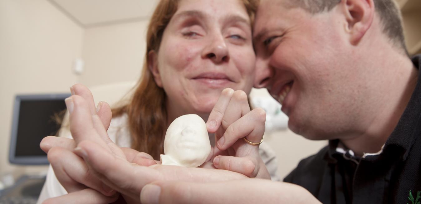
Dr. Jean-Marc Levaillant knows how special it is for expecting parents to see their baby for the first time on an ultrasound. But helping a visually impaired parent “see” the images with a 3D-printed model of their child was even more emotional than the obstetrician anticipated. “She was incredibly moved, and so were we,” Levaillant says. “My whole team was in tears.”
Both Ana Paula and Alvaro, who live in São Paulo, Brazil, are legally blind. Their son, Davi Lucas, was strong and healthy, but there was no way their eyes could see the first grainy glimpses of their baby on the ultrasound monitor.
Whenever Dr. Ralf Menkhaus prepares to administer future parents their first fetal ultrasound, he knows the pressure is on: Equal parts thrilled and anxious, expectant parents are desperate to catch a glimpse of their unborn child. Yet, as a fetal medicine specialist, Menkhaus’ top priority is capturing crucial information about the health of the fetus. He’s looking for evidence, for instance, of conditions like spina bifida, a neural tube defect that affects the spinal cord.
Arzt performs heart surgery on unborn babies, inserting a needle into the mother’s womb and carefully pushing it through a tiny valve in the fetus’ heart that’s just 2 millimeters in diameter, or about as wide as a pinhead. Then he perforates the valve. “If I go 1 or 2 millimeters too far, I tear off the vessel and everything is over,” he says from his office at Kepler University Hospital in Austria, where as head of prenatal care he has overseen more than 140 such procedures.


